Scott M. Goodwin, OMS-4, ENS, MC, USNR
West Virginia School of Osteopathic Medicine
Case
A 26-year-old female, G1P1, presents to the ED with right flank pain of three hours duration. The pain radiates to the right lower quadrant and is intermittent and sharp in nature. The pain is associated with nausea and two episodes of vomiting. However, the patient denies any fever or chills. She denies any dysuria, vaginal bleeding, trauma or any possibility of being pregnant. The patient has no past medical history including any previous abdominal surgeries and admitted only to the use of a contraceptive implant. She denies the use of tobacco and illicit drugs, but admits to the use of ethanol on occasion. On physical examination, the patient appears to be in moderate distress and has reproducible right flank pain. Initial labs including CBC, CMP, and B-hCG are unremarkable with the exception of microhematuria on UA. The findings of a bedside ultrasound are displayed in figures 1.1-1.2
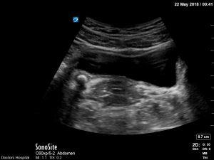
Figure 1.1
Figure 1.1[1] Demonstrates the suprapubic transverse view of a curve-linear probe in a 26-year-old female with acute onset flank pain. Notice the highly echogenic structure on the left side of the image representing an 8.7cm distal ureteral calculus (thin arrow) just proximal to the bladder (star). Notice also the echogenic shadowing (thick arrow) of the associated stone located just lateral to the uterus(circle).
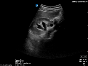
Figure 1.2
Figure 1.2[2] Demonstrates the short axis view of a curve-linear probe in the mid-axial line of a 26-year-old female with acute onset of flank pain. Notice the dilation of the renal collecting system or hydronephrosis (arrow) associated with an obstructing ureteral calculus.
Discussion
In previous years, ultrasound has merely been an ancillary imaging study for most ED physicians. However, with the development of fellowships across the country, ultrasound is quickly becoming standard of care amongst the emergency medicine community, particularly as an initial diagnostic modality. Advantages include availability at the bedside, low cost, no radiation exposure, and no known side effects. Major disadvantages include user reliability. As related to nephrolithiasis, ultrasound has its highest utility in detecting complications of stones such as hydronephrosis (sensitivity 78% for stones < 5mm and 90% for stones >5mm). It is also successful at detecting large stones as well as stones in the proximal and distal ureter. There are limitations visualizing small stones as well as stones in the middle third of the ureter. [2]
One study which compared ultrasound performed by emergency medicine physicians to a standard dose abdominopelvic CT scan demonstrated a 54% sensitivity of ultrasound to detect stones vs. an 88% sensitivity of CT scan. However, the rate of importantly missed diagnoses resulting in complications was comparable between the two groups and the cumulative radiation exposure after six months was approximately 70 percent higher with initial CT. [3]
Ultimately, the utility of ultrasound is in its ability to rapidly detect pathology and assist physicians in early clinical decision making. Although ultrasound does have limitations compared to CT scan, when used as a screening tool in conjunction with CT imaging, it can be a safe and cost-effective tool for emergency physicians. Specifically, with regards to renal calculi, ultrasound should be first utilized to inspect for hydronephrosis and large stones in the proximal and distal ureters, and be used with the understanding that further testing with CT scans may be warranted due to its limited ability to detect smaller stones and other subtle findings.
When considering the disposition of patients with nephrolithiasis, it imperative for the emergency physician to consider the absolute indications for admission including: signs of AKI and sepsis, intractable pain and vomiting, single or transplanted kidney, advanced age and hypercalcemic crisis. [4]
Disposition
In the ED, the patient receives 1L of Normal Saline and a CT scan is not performed. Additionally, she is given IV pain medication including 30mg Toradol and 50mcg Fentanyl as well as 4mg Zofran IV for her nausea. Upon reevaluation, in the ED the patient is pain-free and capable of tolerating PO and is discharged home with oral Norco and a urology follow-up. Appropriate return precautions are addressed including the development of fever, continued or worsening vomiting and pain.
References
1.Morales-Torres, J. Doctors Hospital Images. Columbus, OH. May 2018.
2.Manthey, D. Nicks, B. Urologic Stone Disease: Tintinalli’s Emergency Medicine A Comprehensive Study Guide. 8th Edition. McGaw-Hill Education; 2016. Page 611
3.Curhan, G. Aronson, M. Preminger, G. (2018). Diagnosis and Acute Management of Nephrolithiasis in adults. Lam, A. Lee, S. UpToDate. Retrieved Oct 3, 2018 from https://www.uptodate.com/contents/diagnosis-and-acute-management-of-suspected-nephrolithiasis-in-adults?search=Ultrasound%20emergency%20medicine%20kidney%20stones&source=search_result&selectedTitle=1~150&usage_type=default&display_rank=1
4.Manthey, D. Nicks, B. Urologic Stone Disease: Tintinalli’s Emergency Medicine A Comprehensive Study Guide. 8th Edition. McGaw-Hill Education; 2016. Page 613